The Worlds Within Our Body
In Life Magazine on 9 January 1970, Lennart showed his first images taken using a scanning electron microscope. These were also the first SEM images coloured by Gillis Häägg.

Along the way he assisted scientists with their own research, participated in scientific papers – and pioneered a variety of photographic techniques
Life Magazine, 9 January 1970
-
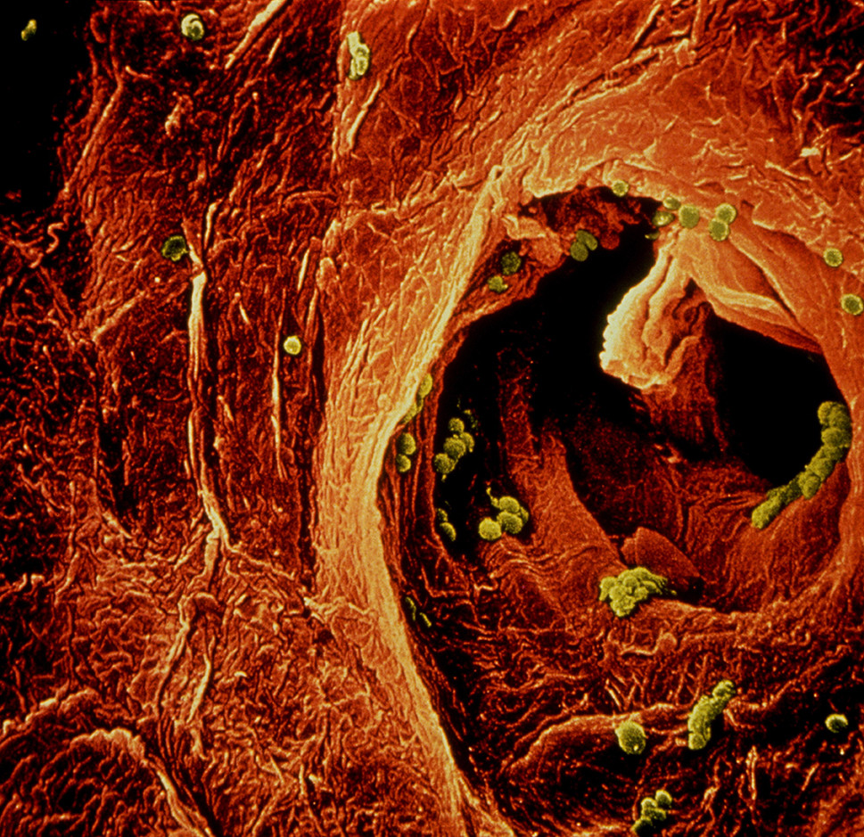
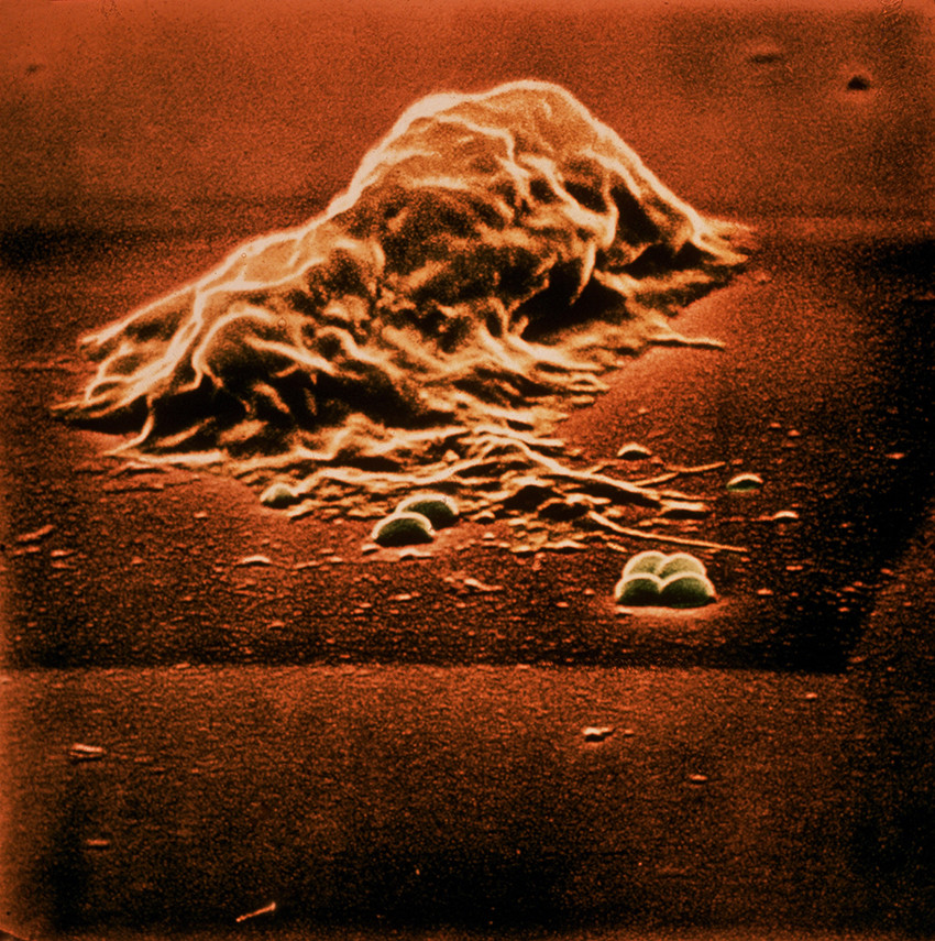
An opening in the fingertip showing sweat emerging. The green “spheres” are bacteria, 1969. ©Lennart Nilsson/SPL -
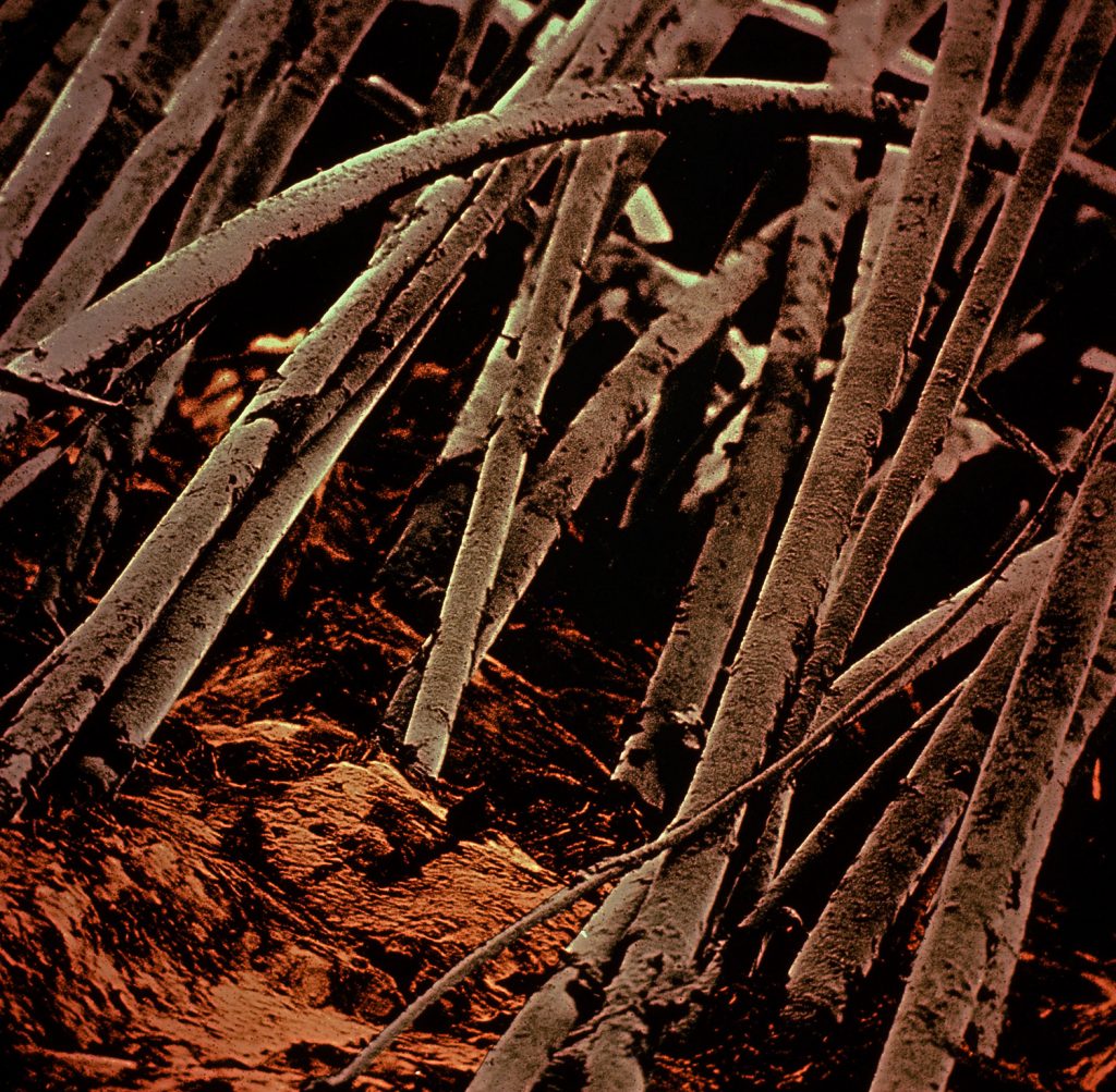
Hair on the head. The first images using scanning electron microscopy, 1969. Coloured by Gillis Häägg the same year. ©Lennart Nilsson/SPL -
Gillis Hääg, 1970 -
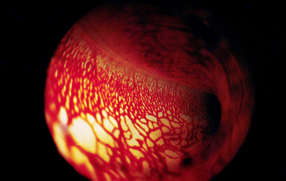
In the cochlear staircase of the inner ear, 1969. ©Lennart Nilsson/SPL -
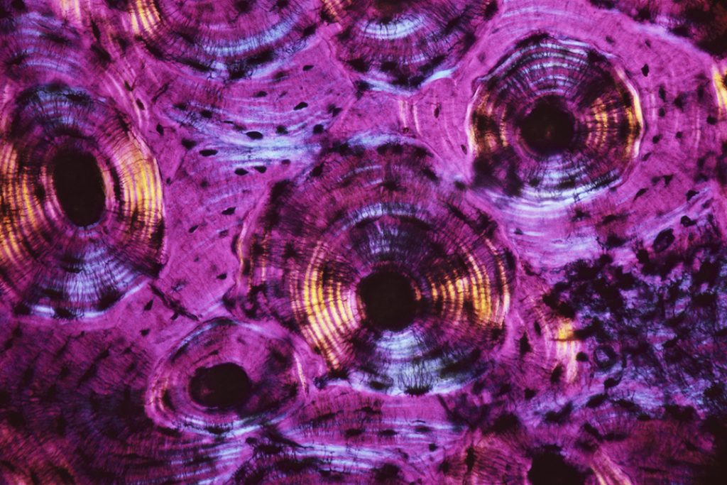
Bone tissue in a microscopic cross-section, 1969. ©Lennart Nilsson/SPL -
The battle between a white blood cell (lymphocyte) and bacteria, 1969. ©Lennart Nilsson/SPL -
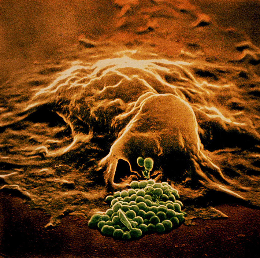
The battle between a white blood cell (lymphocyte) and bacteria, 1969. ©Lennart Nilssson/SPL -
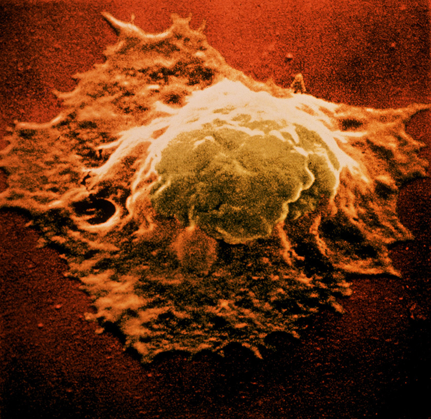
The battle between a white blood cell (lymphocyte) and bacteria, 1969. ©Lennart Nilsson/SPL -
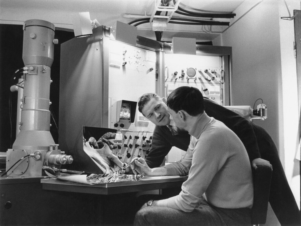
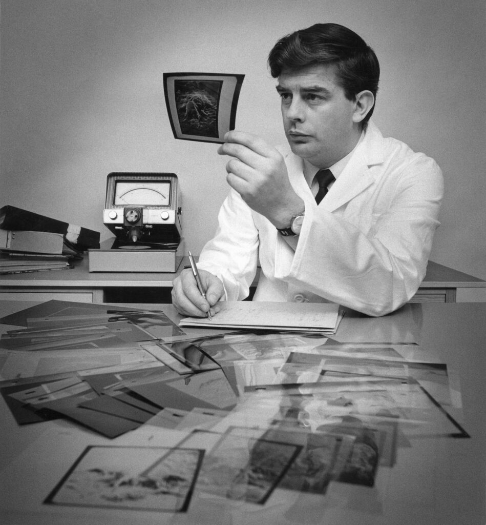
Lennart Nilsson and Göran Alsterborg at Analytica in Stockholm, 1969.


-
An opening in the fingertip showing sweat emerging. The green “spheres” are bacteria, 1969. ©Lennart Nilsson/SPL -
Hair on the head. The first images using scanning electron microscopy, 1969. Coloured by Gillis Häägg the same year. ©Lennart Nilsson/SPL -
Gillis Hääg, 1970 -
In the cochlear staircase of the inner ear, 1969. ©Lennart Nilsson/SPL -
Bone tissue in a microscopic cross-section, 1969. ©Lennart Nilsson/SPL -
The battle between a white blood cell (lymphocyte) and bacteria, 1969. ©Lennart Nilsson/SPL
-
The battle between a white blood cell (lymphocyte) and bacteria, 1969. ©Lennart Nilssson/SPL -
The battle between a white blood cell (lymphocyte) and bacteria, 1969. ©Lennart Nilsson/SPL -
Lennart Nilsson and Göran Alsterborg at Analytica in Stockholm, 1969.
“This extraordinary series of electron microscope pictures shows a large white cell known as a macrophage actually consuming dangerous invaders, staphylococci bacteria. To make these pictures, (the last three images), Photographer Nilsson scraped a few white cells from his own throat, obtained bacteria from Swedish bacteriologists and put them together under a standard light microscope – with a supply of penicillin on hand in case of accidental infection. As each white cell reached a particular stage in its attack on the bacteria, he stopped the process with a fixative chemical and began to job of preparing the specimen for the electron microscope – washing, cleaning and coating it with a delicate gold film designed to reflect electrons and, ultimately, produce the cell’s image on a phosphorescent screen. Nilsson then photographed the screen and, with the help of fellow researchers, added color that matched the original cells as closely as possible. The final sequence provides a remarkable record of the kind of crucial battle that goes on constantly – and invisibly – in our bodies.”
See the feature in Life Magazine


