Verk
Embryonic Studies
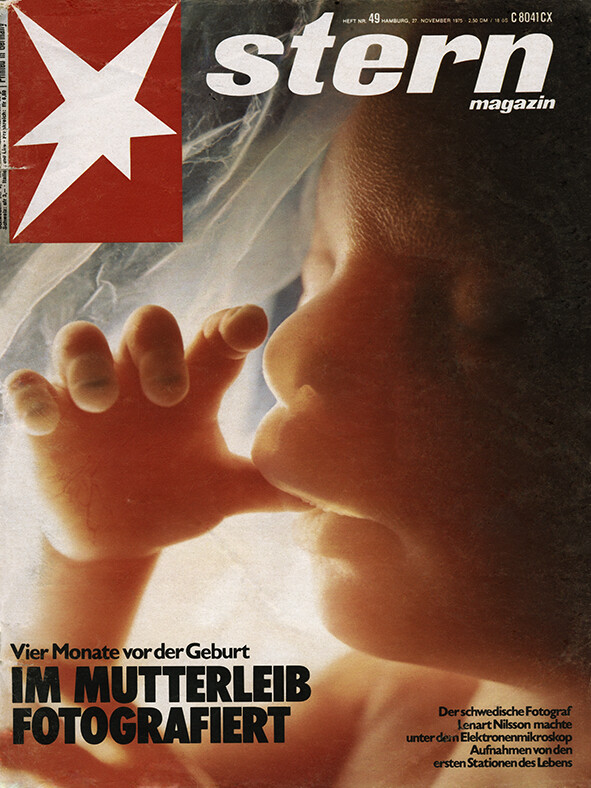
Forensic pathologist Jan Lindberg and photographer Lennart Nilsson present a series of images from studies of around thirty human embryos, the youngest approximately four weeks old and the oldest approximately ten weeks old.
-
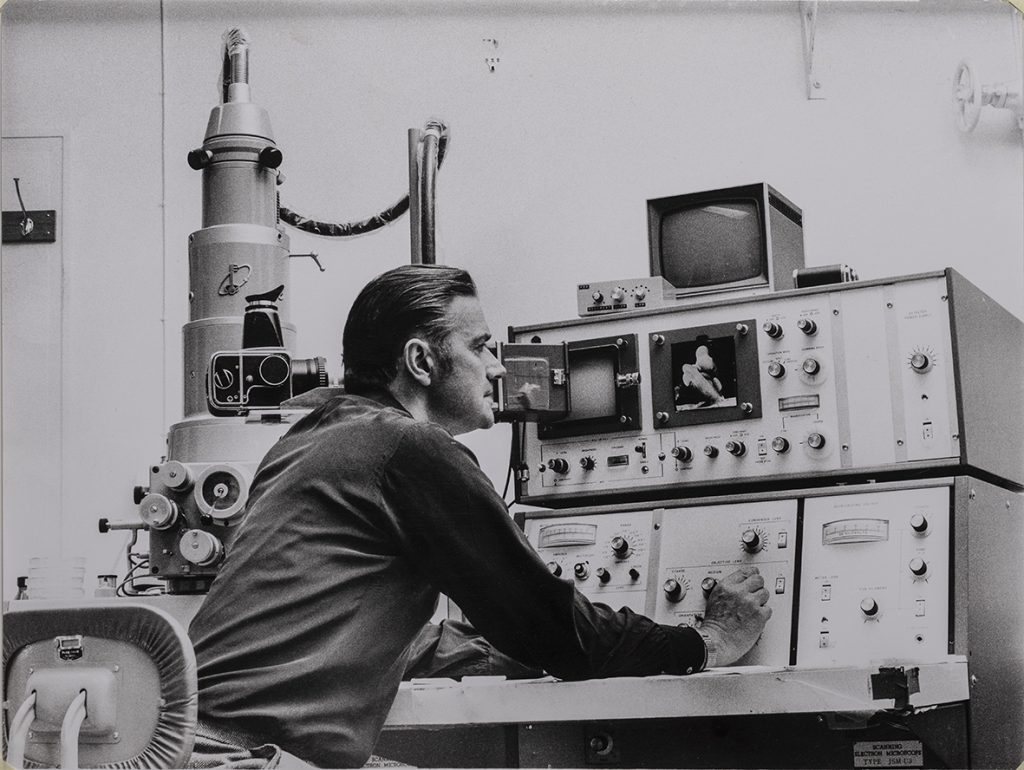
Lennart Nilsson at the scanning electron microscope at the Karolinska Institutet in 1974. © Lennart Nilsson/SPL/TT -
Embryo 5 weeks -
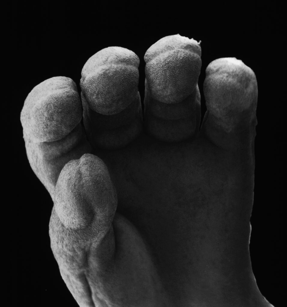
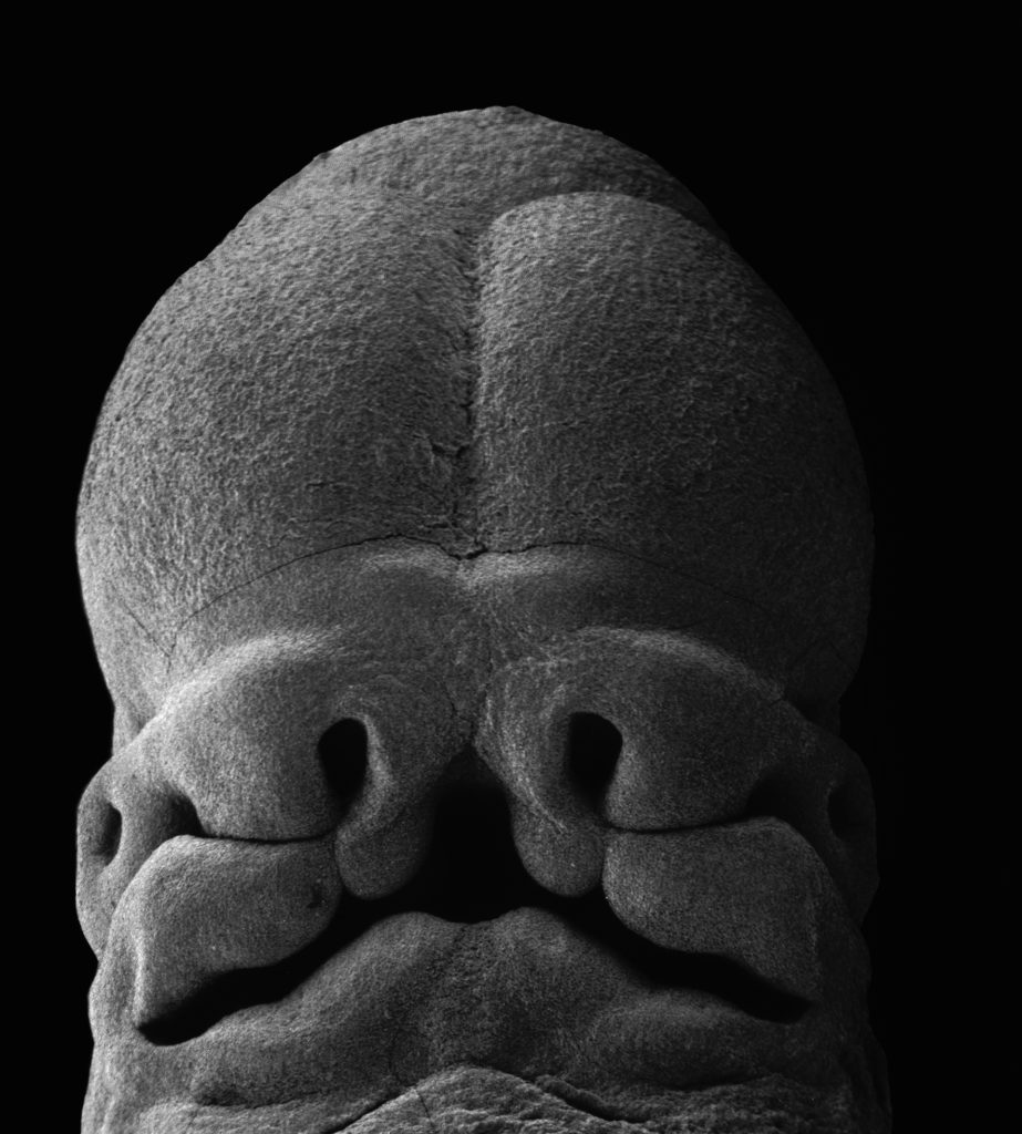
The fingers of the embryo at 11 weeks -
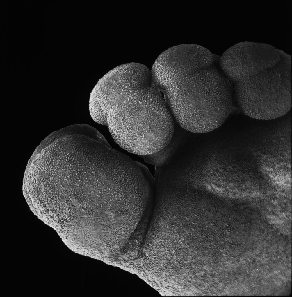
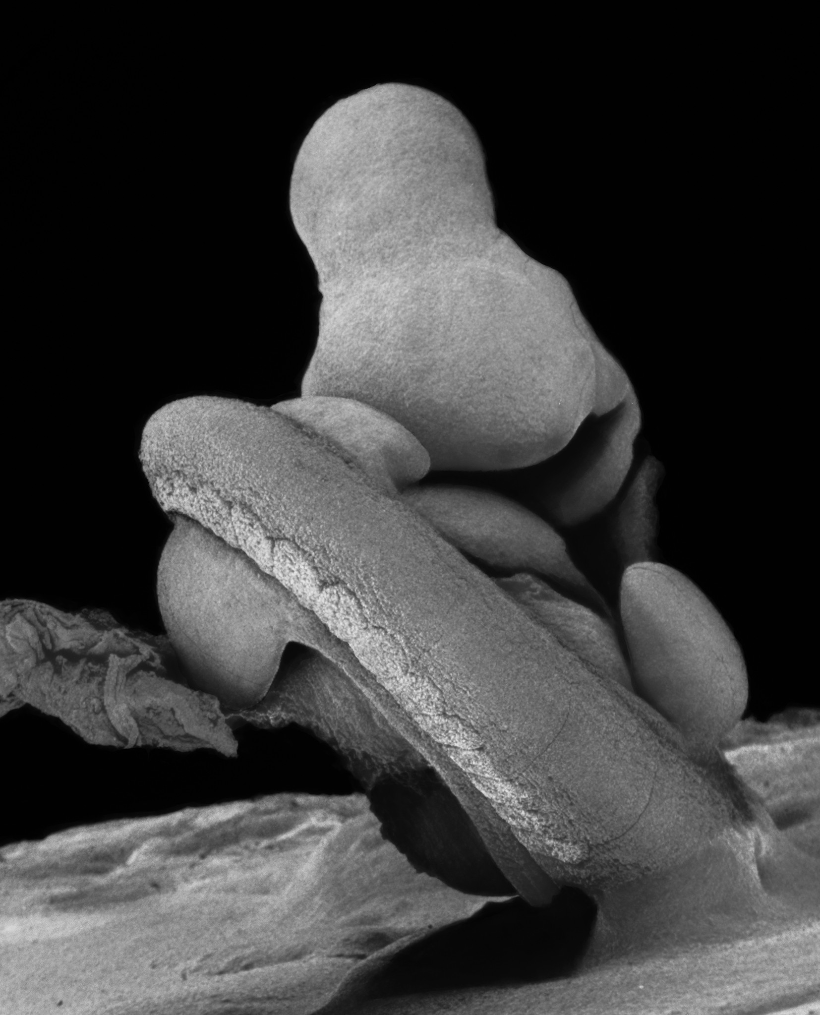
The embryos foot at 11 weeks


-
Lennart Nilsson at the scanning electron microscope at the Karolinska Institutet in 1974. © Lennart Nilsson/SPL/TT -
Embryo 5 weeks -
The fingers of the embryo at 11 weeks -
The embryos foot at 11 weeks
“At the Department of Forensic Medicine at the Karolinska Institutet, we have long studied the earliest stages of human development. Around thirty human embryos of varying ages have been carefully examined in the Japanese scanning electron microscope JEOL type JSM-U3 and photographed using a Hasselblad camera specially mounted in front of the microscope’s display screen, equipped with lens accessories for close-range imaging of objects.”
Läkartidningen (The Swedish Medical Journal) 35/1974