Verk

SEM

Se Människan|1973|Teknik, Vetenskapsfotografi

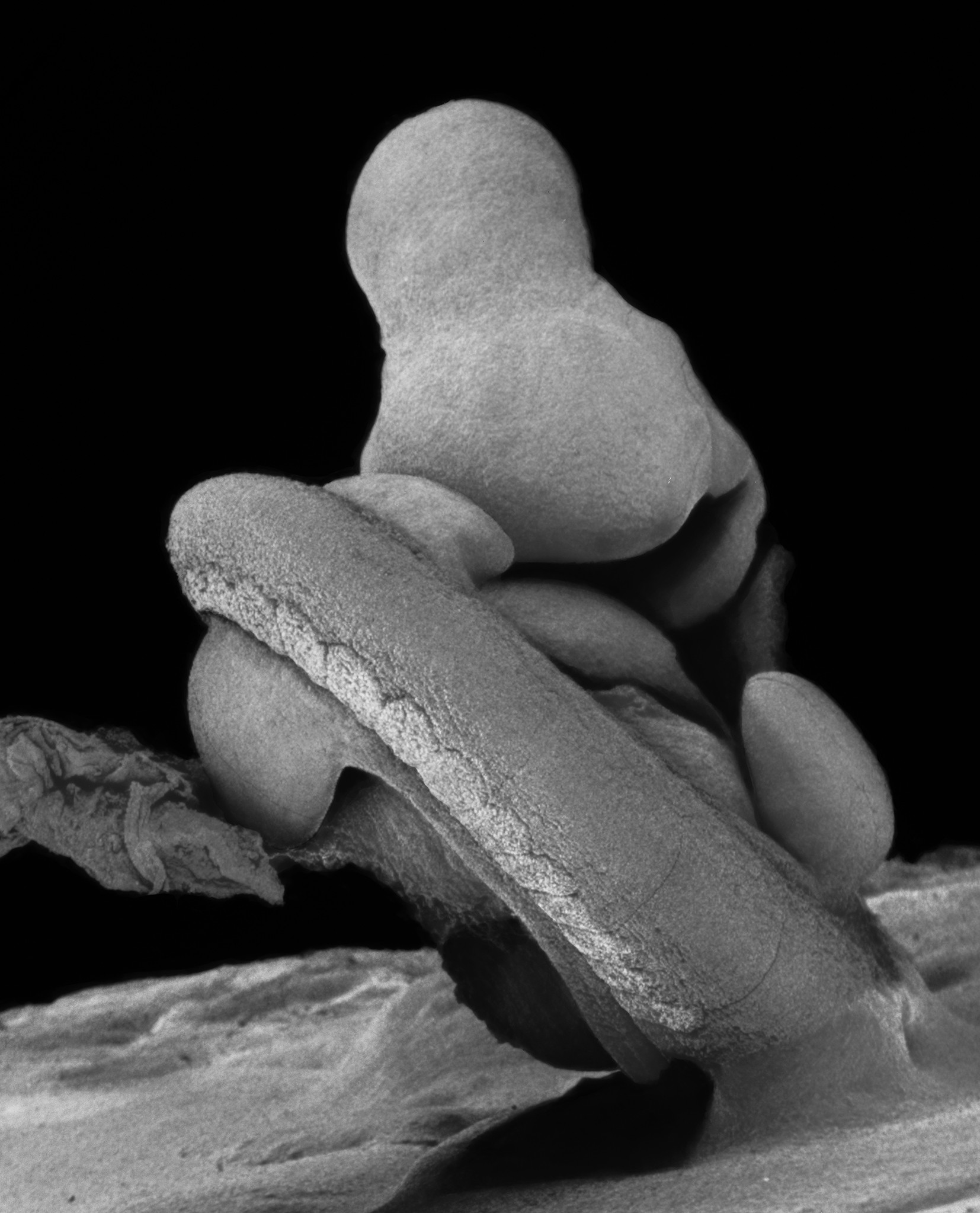
This image of the gastric mucosa was one of the first taken using a scanning electron microscope manufactured by JEOL (JSM-U3). It was Lennart’s first personal SEM and was installed in his laboratory at the Karolinska Institutet in Stockholm. Earlier images, starting in 1969, were taken with the assistance of Göran Alsterborg using the Cambridge scanning electron microscope at the company Analytica in Stockholm.

The stomach contains three different types of glands. The image shows the gastric mucosa in the lower part of the stomach, near the lower stomach opening, the pylorus. Here, so-called pyloric glands are found. These are simple glands that secrete mucus. Near the upper stomach opening, there are also mucus-producing glands. In the largest part of the stomach, there are so-called fundic glands. They secrete enzymes and hydrochloric acid. (SEM)