Verk
Macro and micro
Nilsson took his first images through a microscope already as a teenager. In the early 1950s, he published a study on the contents of water and another on chlorophyll using this technique. He received help from Harry Kinnander at Stockholm University, an important figure in Lennart’s knowledge of microscopy. Around the same time, he also began taking his first macro photographs of subjects from the animal world. It was during this period, in 1952, that he took his first photograph of an embryo.

It was Harry Kinnander who had the patience to teach me microscopy, and for that I am forever grateful to him.
Lennart Nilsson
-
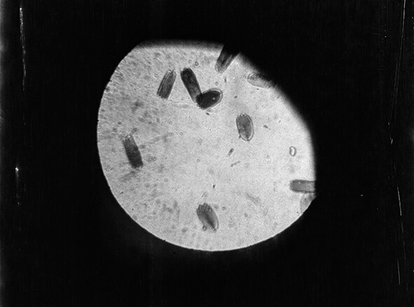
The first image taken through a microscope around 1938. -
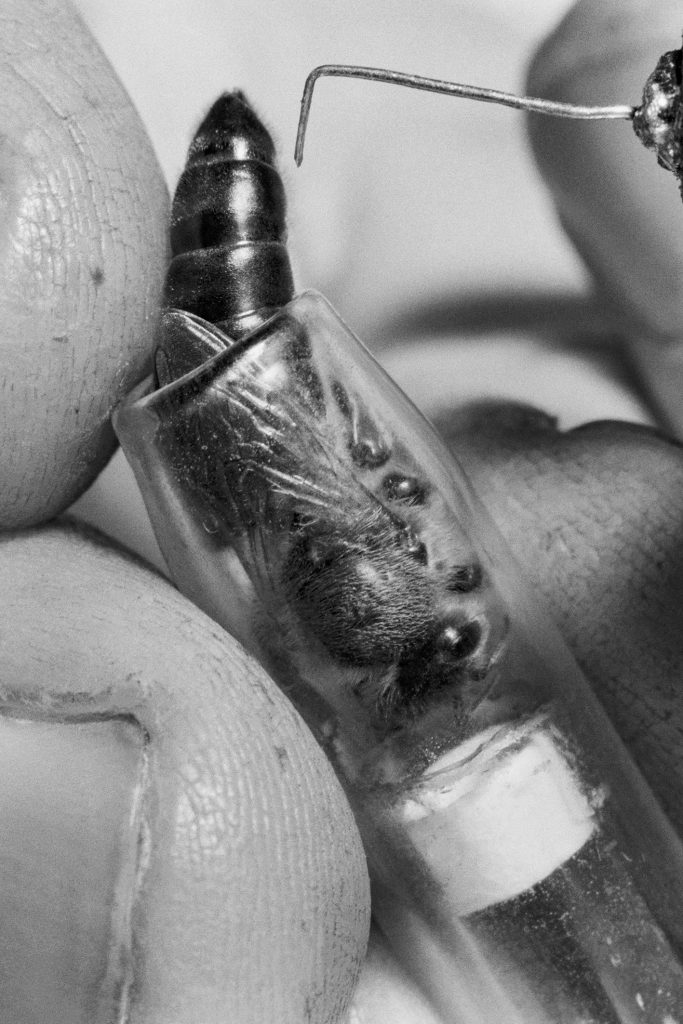
“Only a few days old, the queen bee is ready for fertilization. Instead of a dizzying ascent into the sky with suitors in tow, she is gently placed in a glass tube.” The Queen Bee’s Test-Tube Offspring, 1951. -
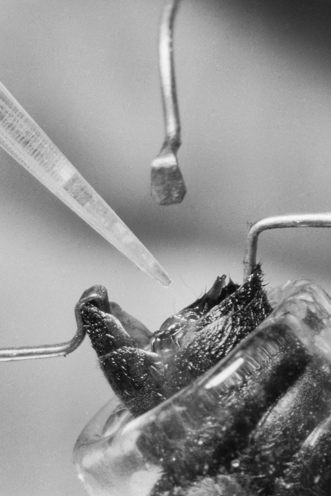
“Semen from at least three drones—1 cubic millimeter from each—is used in the artificial insemination of the queen bee. The sperm cells remain alive and suffice for the queen’s entire lifetime.” The Queen Bee’s Test-Tube Offspring, 1951. -
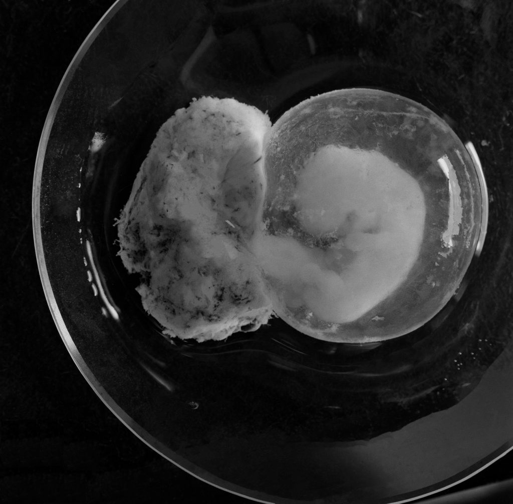
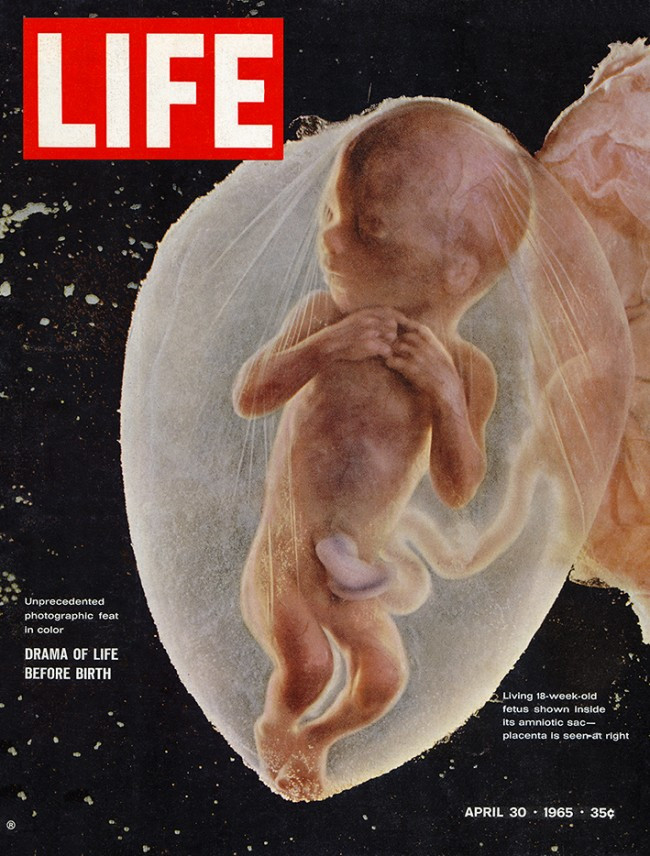
Embryo in formalin, Stockholm, 1952. ©Lennart Nilsson/SPL -
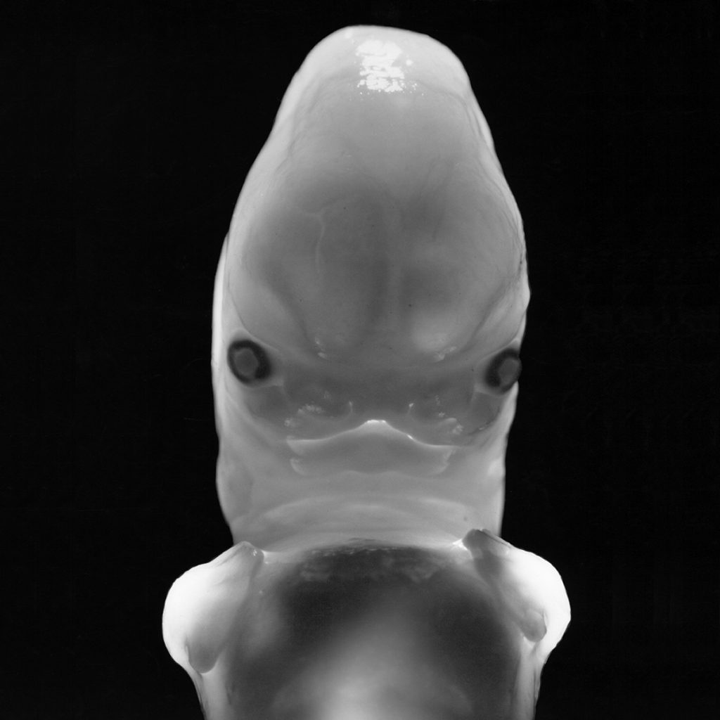
Embryo, 6 weeks, Stockholm 1952 ©Lennart Nilsson/SPL -
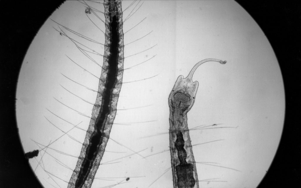
Common freshwater worm with a tentacle-like, extended head lobe. The worm crawls among aquatic plants using its bristles, 1952. -
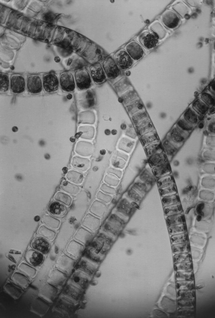
A few filaments of a green alga, forming a green fringe just below the waterline in many of our lakes, 1952. ©Lennart Nilsson/SPL -
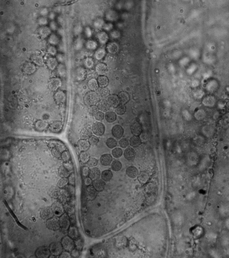
A cell from a leaf of ‘Elodea’ (waterweed). Inside the cell are numerous round bodies, chloroplasts, which carry the chlorophyll, 1953. ©Lennart Nilsson Photography/SPL


-
The first image taken through a microscope around 1938. -
“Only a few days old, the queen bee is ready for fertilization. Instead of a dizzying ascent into the sky with suitors in tow, she is gently placed in a glass tube.” The Queen Bee’s Test-Tube Offspring, 1951. -
“Semen from at least three drones—1 cubic millimeter from each—is used in the artificial insemination of the queen bee. The sperm cells remain alive and suffice for the queen’s entire lifetime.” The Queen Bee’s Test-Tube Offspring, 1951. -
Embryo in formalin, Stockholm, 1952. ©Lennart Nilsson/SPL -
Embryo, 6 weeks, Stockholm 1952 ©Lennart Nilsson/SPL -
Common freshwater worm with a tentacle-like, extended head lobe. The worm crawls among aquatic plants using its bristles, 1952.
-
A few filaments of a green alga, forming a green fringe just below the waterline in many of our lakes, 1952. ©Lennart Nilsson/SPL -
A cell from a leaf of ‘Elodea’ (waterweed). Inside the cell are numerous round bodies, chloroplasts, which carry the chlorophyll, 1953. ©Lennart Nilsson Photography/SPL